
A 3D scan captures far more information than a traditional X-ray, showing the exact position of your teeth and jawbone in relation to nearby nerves and sinuses. This level of detail helps our team plan procedures like implants or extractions with precision. It reduces surprises and ensures reliable outcomes.
3D Cone Beam Imaging (CBCT)
Enjoy Advanced Smile Care
Get Precise Care With 3D Cone Beam Imaging in Montgomery
Seeing beyond what a flat X-ray shows makes a real difference in how your dental treatment gets planned. 3D cone beam imaging in Montgomery captures a precise view of your teeth and jawbone in a single scan, revealing detail that isn't possible with a regular X-ray.
3D imaging gives your dentist a clear picture of your nerves and bone density before starting treatment. It plays an important role in planning dental implants and other more involved procedures. The scan takes only a few seconds and is completely comfortable.
Discover How 3D Imaging Technology Improves Your Care
Superior Detail
Unlike a flat X-ray, this advanced scanning technology creates a 3D image that shows depth and angles that a standard 2D picture can't capture.
Used Across Treatments
3D imaging helps your dentist plan several types of treatment, from implants to oral surgery, making it a versatile part of your overall care.
Precise Treatment Planning
Seeing the exact position of nerves and bone density beforehand helps your dentist plan procedures like implants with added precision.
Quick, Comfortable Scan
The cone beam scan itself takes only seconds and requires no injections or preparation. You just remain still for a few moments.
Find Out What's Involved in 3D Scanning
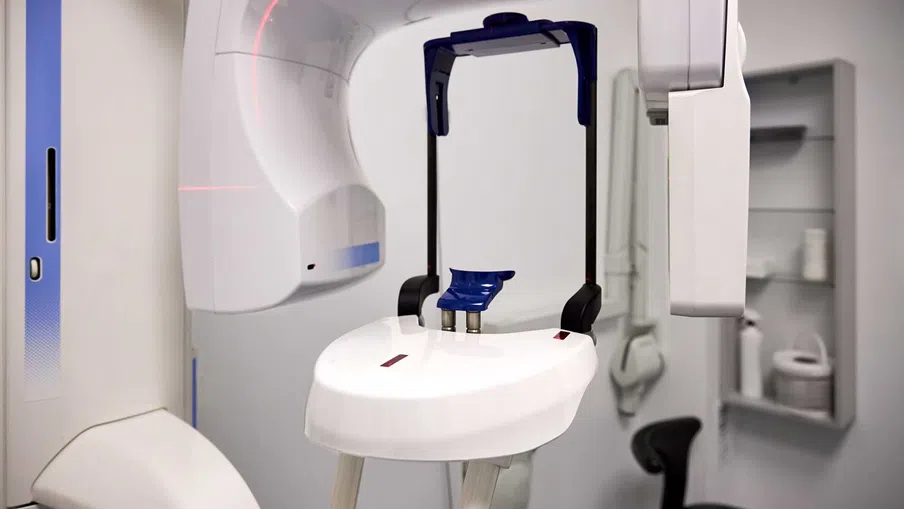
Taking a 3D scan involves sitting in a chair while the machine rotates slowly around your head, capturing images from multiple angles in just a few seconds. No special preparation is needed beforehand, and the process feels similar to a standard dental X-ray taken at a checkup.
Our team uses 3D cone bean CT imaging in several different ways:
- Planning implant placement around nerves and jawbone density
- Evaluating a tooth before a root canal or extraction
- Assessing the jaw and airway for sleep apnea concerns
- Mapping bone before a graft or other oral surgery procedure
Since the scan is completed in the office during the same visit as a consultation, results are typically available right away to help plan your next steps.
If you'd like to learn more about 3D cone beam imaging in Montgomery, contact our office today.
Frequently Asked Questions About 3D Cone Beam Imaging
Q What is 3D cone beam imaging?
A
3D cone beam imaging, often called CBCT, is a type of dental X-ray that captures a detailed view of the teeth, jawbone, and surrounding structures. The machine rotates around the head, taking multiple images that combine into a detailed 3D picture.
Q How is CBCT different from a regular dental X-ray?
A
A regular dental X-ray captures a flat, 2D image, while a CBCT scan produces a 3D view showing depth and spatial relationships. This added detail helps a dentist see things a standard X-ray might miss, particularly around bone and nerve positions.
Q Is 3D cone beam imaging safe?
A
3D cone beam imaging is considered safe for most patients. It uses a low dose of radiation similar to or sometimes less than a full series of traditional dental X-rays. The equipment is designed specifically to minimize exposure while still capturing detailed images.
Q How much radiation is involved in a CBCT scan?
A
The radiation involved in a CBCT scan is generally low, though the exact amount depends on the specific machine and settings used for the scan. Modern equipment is designed to capture detailed images while keeping radiation exposure as limited as reasonably possible.
Q How long does a 3D scan take?
A
A 3D scan typically takes only a few seconds to complete, though the entire appointment, including positioning, may take a few minutes total. This is often quicker than a full series of traditional X-rays covering the same areas.
Q Does a CBCT scan hurt?
A
A CBCT scan doesn't hurt, since it simply involves sitting still while a machine captures images from outside the mouth. There's no need for anesthesia, injections, or any physical contact with the teeth or gums during the scan.
Q What is a 3D scan used for?
A
A 3D scan is commonly used to plan dental implants, evaluate teeth before a root canal or extraction, and assess the jaw for sleep apnea concerns. It also helps plan bone grafts and other procedures involving the jawbone or surrounding structures.
Q How does 3D imaging compare to a traditional panoramic X-ray?
A
A panoramic X-ray captures a wide, flat image of the entire mouth in one shot, while a CBCT scan creates a 3D model from multiple angles. Panoramic X-rays are often used for a general overview, while CBCT provides more precise detail for specific procedures.


