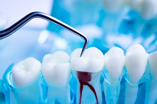
Root Canal When the soft tissues in the pulp of the tooth become infected or inflamed, they need to be removed with a root canal. The pulp is the soft core of the tooth. When bacteria invades the pulp it can agitate the nerve causing severe tooth pain and in some cases form an abscess at the root of the tooth. Tooth decay can be caused by poor dental hygiene and repeated dental procedures on the tooth. Chips or cracks in the enamel from force trauma can allow harmful bacteria into the tooth. Injury or force trauma to the tooth can damage the root leading to inflammation from the inside out even if there is no visible decay, chips, or cracks on the outside of the tooth. Symptoms of tooth infection include: lingering pain in a tooth when biting or chewing, pimples on the gums, lingering tooth sensitivity to temperature, deep decay or darkening of the gums, chipped or cracked teeth, swollen or tender gums. Scheduling a dental exam with one of our dentists will assess your risk of infection, or need for a root canal. Schedule an appointment with Masci, Hale & Wilson Advanced Aesthetic and Restorative Dentistry today. When the soft tissues in the pulp of the tooth become infected or inflamed, they need to be removed with a root canal. The pulp is the soft core of the tooth. When bacteria invades the pulp it can agitate the nerve causing severe tooth pain and in some cases form an abscess at the root of the tooth. Tooth decay can be caused by poor dental hygiene and repeated dental procedures on the tooth. Chips or cracks in the enamel from force trauma can allow harmful bacteria into the tooth. Injury or force trauma to the tooth can damage the root leading to inflammation from the inside out even if there is no visible decay, chips, or cracks on the outside of the tooth. Symptoms of tooth infection include: lingering pain in a tooth when biting or chewing, pimples on the gums, lingering tooth sensitivity to temperature, deep decay or darkening of the gums, chipped or cracked teeth, swollen or tender gums. Scheduling a dental exam with one of our dentists will assess your risk of infection, or need for a root canal. Schedule an appointment with Masci, Hale & Wilson Advanced Aesthetic and Restorative Dentistry today.Anatomy Of The ToothUnderstanding the necessity for root canal starts with understanding tooth anatomy. The top outer layer of the tooth is the enamel. It's a protective layer, the part that you see when you smile. Beneath it is a hard layer called dentin; it forms most of the tooth's body. Inside the dentin is a chamber filled with soft tissue called the pulp. The pulp is fleshy, composed mostly of blood vessels and nerve endings. The pulp forms the surrounding hard tissues of the tooth during development by providing it with nutrients. When the tooth fully matures, it can survive without its pulp, taking nutrients from surrounding tissues. Root Canal ProcedureRoot canal procedures can span over two or more dental visits. Consider scheduling a consultation with one of our dentists at Masci, Hale & Wilson Advanced Aesthetic and Restorative Dentistry to learn more about the procedure and form a plan for treatment. To start the procedure, an image of the tooth and its supporting structure is taken using an x-ray. The image is used to assess the level of infection and areas of infection in the tooth. Ruling out the cause of infection helps the dentist administer the correct treatment. After the imaging, an anesthetic is administered, to numb the tooth and surrounding tissue. During the root canal, the tooth needs to be kept dry, so a small protective sheet called a dental dam is placed around the tooth, isolating it from neighboring teeth and saliva. Now, a small hole is drilled in the tooth using a dental drill. Using small thin instruments, the infected pulp is removed from the core and root of the tooth. The now-vacant space is cleaned and shaped and the root is plugged with a biocompatible material. In most cases the dentist will place a filling over the chamber, sealing the tooth from further infection. After this stage, your dentist will have you come back to remove the filling and place a crown over the tooth. If you are considering a root canal, schedule a dental exam today at (845) 457-5763. |
HoursMon - Fri: 7:30am - 5:00pm Closed Saturday & Sunday |